Sprain of the ligaments of the ankle is the most common ankle injury. The ankle is capable of tolerating tremendous force. It has been estimated that during a run the ankle bears 9-13 times body weight at peak force.
How do you know if you sprained your ankle?
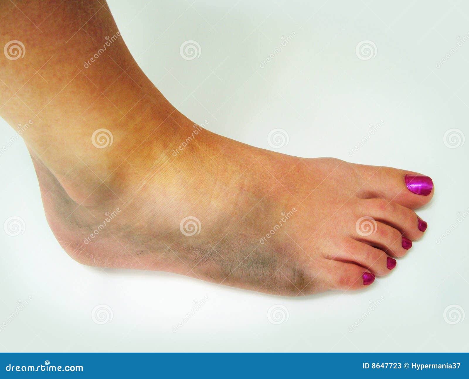
The signs and symptoms of an ankle sprain include pain, swelling and discoloration of the ankle.
So you think you just sprained your ankle, what should you do? Many people underestimate their ankle sprain and end up ignoring it. What if you leave an Ankle Sprain untreated? Did you know that by putting up with the pain and swelling for too long you may be further injuring your ankle? If an ankle sprain is not diagnosed or treated properly the ligaments stretch out leading to further injury. It is estimated that up to 70% of ankle sprains result in longterm pain and disability. Repeated ankle sprains can lead to Chronic Ankle Instability (CAI) and Osteoarthritis. Clinical guidelines have been developed to insure that all ankle sprains are diagnosed and treated correctly.
What is Chronic Ankle Instability?
Chronic Ankle Instability (CAI) is a stretching or weakness of the ankle ligaments such that rather than supporting the ankle, the ankle “gives out” or “rolls” easily. CAI may be caused by repeated ankle sprains or an ankle sprain which was not treated and never healed properly.
Call For An Appointment: (814) 882-2663
What’s the Best Way to Treat an Ankle Sprain?
Who among us has not sprained an ankle or a ligament? These injuries are extremely common both in children and adults. According to the authors at UpToDate, ankle sprains are the number one injury seen at the emergency room! People have sprained an ankle while running, jumping or even walking their dog! Uneven surfaces are notorious for causing us to “Roll” our ankles.
Two research studies published in 2019 studied mice with ankle ligament injuries similar to human ankle sprains. They specifically studied the ankle ligaments involved in the most common ankle sprains, the talofibular and calcaneofibular ligaments which are lateral ankle ligaments. The mice ‘rested’ for either 3,7, or 14 days following the ankle ligament injury. In both research studies the mice who rested 14 days performed better in a 45 week follow-up as compared to the 3 or 7 day rest groups. Tricia Hubbard-Turner Ph.D and colleagues found better physical activity results in mice who rested 14 days after ankle injury. Erik Wikstrom Ph.D. and colleagues reported that prolonged rest; longer then 2 weeks, improved dynamic balance in the longterm when tested at 42 and 54 weeks after injury.
What should you do Immediately after an Ankle Sprain
First, Rest. Get off of the injured ankle. Do not walk on it any longer than you have to, do not keep running or cycling. Ice. Apply cold compress or an ice bag, the cold actually helps relieve inflammation. Compression. Apply an ACE bandage or elastic ankle support. Elevation. Prop the leg up at an elevation higher than your chest or just on a chair, by propping it up you reduce swelling.
Types of Ankle Sprains
According to the National Institutes of Health, NIH; http://sph.sagepub.com/ Lateral ankle sprains comprised 91.74%, medial ankle sprains 0.26%, high ankle sprains 0.18%, and “unknown” 7.82% The lateral ankle sprain is by far the most common.
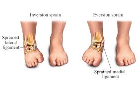
In an Inversion Sprain the Lateral Ligaments are injured. Whereas in an Eversion Sprain the Medial Ligaments are injured.
The problem is that many are never diagnosed nor treated properly.
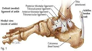
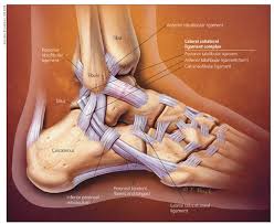
Which Ligaments are injured in an Ankle Sprain?
Please see the images below.
How Long does it take my ankle sprain to Heal? What is the Recovery time?
The time to heal depends upon the Severity or Grade of ankle sprain
Grades of Ankle Sprains and average Healing Time
Grade 1: Mild stretching, no tearing. Recovery time 1-3 weeks.
Grade 2: Partial tear, mild to moderate joint instability. Recovery time 3-6 weeks
Grade 3: Complete tear, loss of function, inability to bear weight. Recovery time several months.
The severity of an Ankle Sprain is dependent upon the number of ligaments injured, the more ligaments injured the more severe the sprain and subsequently, the longer the recovery. Most ankle sprains do not require surgery, however; if there is a Grade 3 complete tear surgery is recommended.
Is it OK to Walk on a Sprained Ankle?
It is best to Rest an injury for two reasons; First, by getting off the injured limb you prevent further damage and Pain, Second, only after a few hours to days can you tell how severe the injury is.
Preventing Further Injury
In order to recover from an ankle sprain properly and prevent further injury and arthritis it is best to seek medical care and rehabilitation. Dr. Danielle Torp at the University of North Carolina at Charlotte, UNCC; led a study where patients with Chronic Ankle Instability were given biofeedback training in order to protect their ankles from further injury. Essentially, patients ‘Learned’ how to walk in a safer manner.
In an article published in July 2018, researchers Drs. Owoeye, Palacios-Derflingher and Emery found that a pre-practice warmup which includes Neuromuscular Training reduced the risk of Ankle Sprain by 32%.
What helps a sprained ankle heal faster?
As with all injures, early diagnosis and treatment results in not only less suffering but a more favorable outcome in the long term. If you get help early you will have a better outcome. The problem with ignoring these injuries is that the ligament will stretch and not heal properly, as a result it will be predisposed to further injury which will lead to even further injury eventually requiring surgery and likely irreversible consequences. Its a domino effect. Ankle sprain if ignored becomes weak. In my office I use diagnostics sensitive enough to reveal a ligament injury before an x-ray or MRI will reveal it. If you’ve sprained your ankle, I can help. Call for your appointment today. 814.882.2663.
About Tendons and Ligaments
Tendons and ligaments are similarly constructed, made of different types of collagen and other factors. Tendons cause joint movement while ligaments restrict joint movement to within a safe range. The types of collagen are I,III,IV, V and VI. Chemicals called proteoglycans function to lubricate and organize collagen fiber bundles. The special feature of tendons and ligaments, their elasticity; is due to type I collagen. The triple helical polypeptide chains are folded and bundled in a hierarchical structure which renders mechanical strength. These fibrils, fibers and fascicles are surrounded by endotenon and epitenon ( a sheath or wrapping) which has its own nerve and blood supply.
About Joints and Joint Fluid
Joints are where ends of bone meet in order to allow movement. The ends of bone are covered by a layer of cartilage and surrounded by synovium. It is the synovium which produces synovial fluid or joint fluid. The synovium reduces friction between the ends of cartilage during movement, hence it functions as a lubricant. Cartilage accepts high mechanical stress and acts as a shock absorber. Cartilage is made of three types of protein: collagen type II, proteoglycans and non-collagenous protein. The interaction between the collagen and proteoglycan is responsible for the compressive and tensile strength of cartilage.